Ultrasound is a very valuable diagnostic tool that we use every day in practice. In this blog, I will explain what ultrasound is and what it can and cannot do. Hopefully after reading this, you will understand the value and limitations of this diagnostic tool so you can understand why we may recommend this for your pet. Most people are familiar with ultrasound mainly as it relates to human pregnancy. It uses sound waves to penetrate soft tissues and these waves are sent back to the ultrasound machine and creates the images we see on the screen. We use our ultrasound machine multiple times a day to get urine samples directly from the urinary bladder. We can visualize the bladder and can actually see a needle go into the bladder to obtain the sample. This is the simplest way we use this piece of technology.
What's the difference between X-rays and Ultrasounds?
Before discussing specifics, it is important to know the difference between an ultrasound and x-rays. We use radiographs (x-ray) to image bones and to get an overview of what a chest or abdomen looks like. We can see size and shape of organs and how they relate to each other. Ultrasound lets us see the structure of the soft tissue organs. They are like two different types of maps, one of a state: The X-ray, having less details and more of an overview, VS a map of a city: The Ultrasound, with more details like the street names present. We use both modalities when we are trying to diagnose any medical concerns. It is important to use both tools, especially in older animals that we are looking into weight loss for example. For example, if we jump right to ultrasound, we are going to possibly miss evidence of cancer in the chest that would be seen if x-rays were taken first.
Why might a doctor suggest an ultrasound for your pet?
Some common reasons we will suggest an abdominal ultrasound would include:
- Elevated liver enzymes on blood work
- Elevated kidney numbers on blood work
- Unexplained weight loss or gastrointestinal signs
- Concerns for an abdominal mass or free fluid seen on radiographs
- Concern for an intestinal obstruction seen on radiographs

What are advantages of an ultrasound?
Ultrasound has many advantages as it is safe and noninvasive. We can see the internal structure of specific organs and we can measure the size of organs and any masses we may find. It is also a great screening tool for when older animals have normal radiographs, but we're still worried something could be going on with them. Ultrasound also can allow us to obtain fine needle aspirate or biopsy samples from an organ or mass in a noninvasive way.
When might a doctor decide against an ultrasound?
There are times where ultrasound is limited and these include very large or obese dogs, dogs with very large masses, or a significant amount of free fluid in the abdomen. In these cases, we may opt for skipping an ultrasound as we know it will not provide a lot of value.
What can't ultrasounds do?
It is really important to understand what ultrasound cannot do; the expectations are aligned with the information we obtain from a study. Ultrasound cannot provide a specific diagnosis for everything. The goal is to form a logical list of possibilities. It cannot determine if something is benign or malignant, although, certain qualities within an image can be suggestive. Ultrasound results also cannot determine function on particular organs. Most importantly, it cannot rule out disease. You can have a normal study, but there is still disease you have not found yet.
What happens during an abdominal ultrasound?
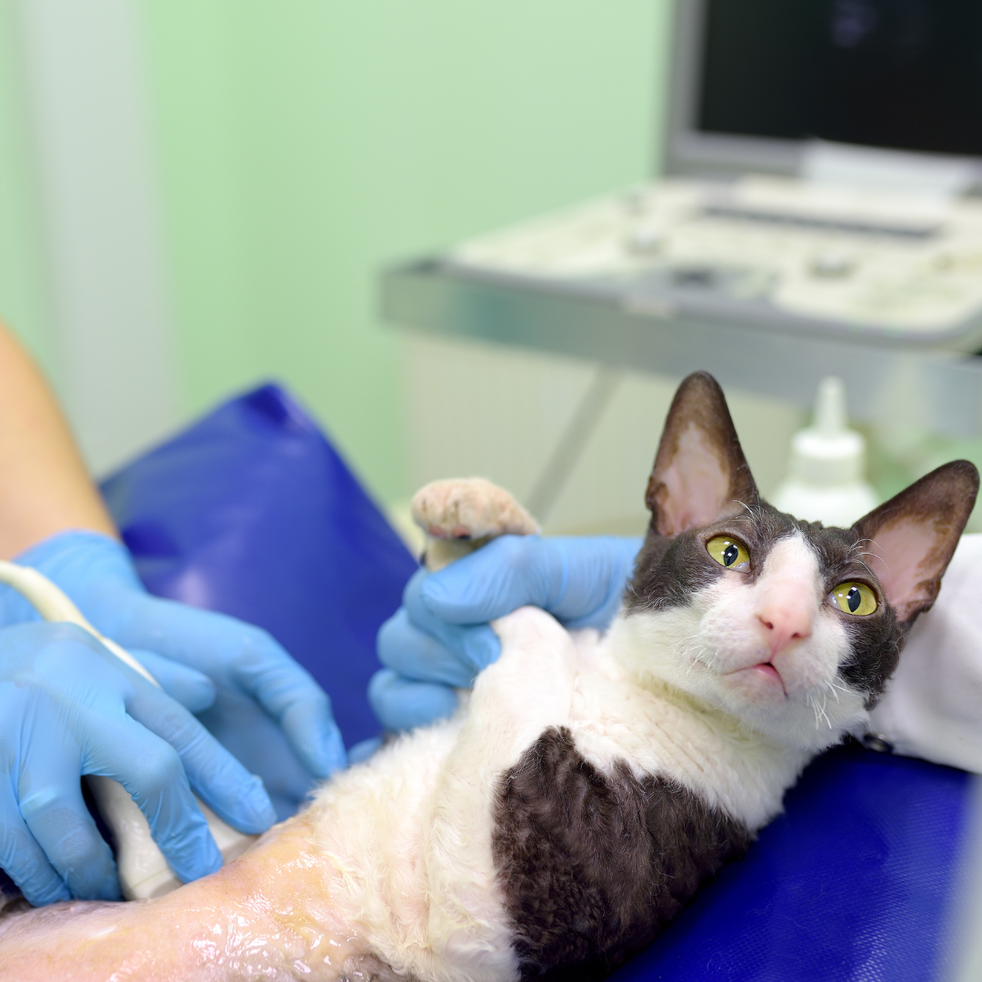
The procedure of an abdominal ultrasound is quite simple. The dog or cat needs to be fasted 8-12 hours prior to the study. Ingested foods filling the GI tract will make the study really difficult and it does not allow you to assess the GI tract properly. Pets need to be shaved along their belly so that the ultrasound probe has the best contact with the skin. We apply an ultrasound gel that allows the sound waves to create the clearest images. Sometimes, we need to use very light sedation to get the best study. Some dogs and cats resist laying on their backs or they are panting a lot, and sedation really helps to make the study efficient and of high diagnostic quality. Once the study is done, we may recommend follow up testing/lab work based on the results of the study.
The Drake Center for Veterinary Care is an AAHA-accredited animal hospital located in Encinitas, CA. The Drake Center loves being a source of information for all pet owners across the country however if you have any questions regarding pet care and do not live in Encinitas, CA or surrounding cities, we encourage you to contact your local veterinarian.
If you have questions and you'd like to reach out to us, you can call us directly at (760) 452-3190, or you can email us at [email protected]. Don't forget to follow us on social media Facebook, Instagram.

